 July 13, 2010 (Peabody, Mass.) – This August, JEOL USA will demonstrate the first correlative microscope to enable concurrent light microscopy and atmospheric scanning electron microscopy (ASEM) for observation of and experimentation on samples in their native state. The new JEOL ClairScope will make its debut in the United States at Microscopy & Microanalysis (M&M) in Portland, Oregon August 2-5, 2010, just prior to installation at Northwestern University’s Biological Imaging Facility where it will be used for demonstrations and applications development.
Acknowledging the significance of this new instrument, the R&D 100 Award, known as the “Oscar of Innovation,” was bestowed on the JEOL ClairScope on July 8 by the editors of R&D Magazine.
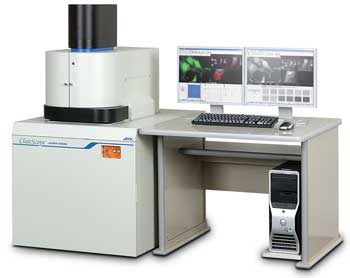
“We’re very excited about introducing this unique microscope to the United States,” Donna Guarrera, Asst. Director of the Scanning Microscopy Division of JEOL USA reports. “I had the opportunity to work with the principal developers of the ClairScope at JEOL headquarters in Japan last month. With just one mouse click, it switches from the optical microscope mode to the ASEM mode. SEM imaging in an open environment will provide a lot of new opportunities to the scientific community.”
Guarrera will be giving one-on-one demonstrations of the ClairScope at M&M 2010, the industry’s largest trade show, in the JEOL booth #636. A ClairScope Workshop will be held from 4:00 – 5:00 p.m., Tuesday, August 2. To preregister or to see a list of related posters to be presented during M&M, visit the Events section of the JEOL USA website at www.jeolusa.com.
Northwestern University to Develop New Applications with ClairScope
JEOL has selected Northwestern University’s Biological Imaging Facility (BIF) as only one of two sites in the world where the ClairScope will be used for applications development. BIF is a research and training facility that serves the imaging needs of over 500 scientists representing 144 different labs, including investigators in the Robert H. Lurie Comprehensive Cancer Center, the Howard Hughes Medical Institute, and the Daniel F. and Ada L.Rice Foundation.
“Making this novel technology available to our users will be of significant benefit to their projects and undoubtedly open new research avenues,” said BIF Manger, Dr. William Russin.
On the other side of the globe, the JEOL ClairScope will also be installed at the University of York (York, England) Department of Biology, where scientists will employ the correlative techniques of fluorescence microscopy and atmospheric SEM in their development of novel probes for biological exploration.
ClairScope Enables High Resolution Observation of Life Science and Materials Experiments As They Occur
Using the ClairScope, life science researchers can harness the powerful imaging capability of the SEM to observe biological processes such as platelet generation, distribution of sugar chains, and microbe growth. Materials scientists will be able to observe and record crystallization, electrochemical reactions, emulsion technology, self-assemblies, and dendrite growth as they occur.
The ClairScope makes it possible for biologists to perform routine fluorescent imaging then examine the specimen at high resolution, high magnification without changing instruments or sample position. The wide-field light microscope with emersion lens is co-axially aligned with the A-SEM column. The specimen dish features an ultrathin SiN film window that allows electron beam transmission while the sample is open to atmospheric pressure. Now scientists can add reagents, drugs, and other substances to the sample in order to perform experiments and observe reactions in both liquid and gas environments.