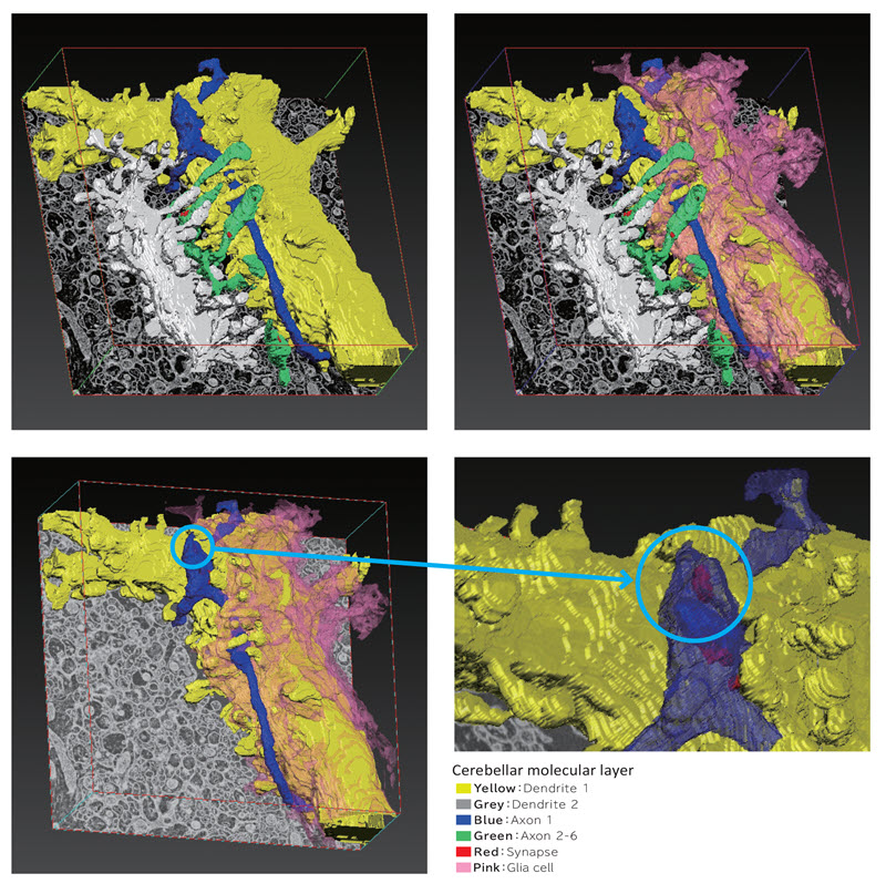
Array tomography provides three-dimensional information of samples such as biological tissues. The sample is fixed and embedded in resin, and is sliced into serial sections. The same positions of the sections are observed using SEM or TEM to make a stack of images, which is used for three dimensional reconstruction.
- High quality images comparable to TEM are available thanks to the improvements of SEM and backscattered electron detector.
- Automatic image acquisition by the newly developed software.
- Observation of large samples is possible.
- Sample preparation of serial sections for SEM is relatively easy, since the sections are placed on solid surface such as silicon wafer or glass slide.
- There is no limitations of sample area and number of slices, allowing large volume three dimensional observation.
- No special equipment is needed, only ultramicrotome and SEM are required tools.
- Serial sections are stained using uranyl acetate and lead citrate, and no special fixation procedure is needed such as NCMIR method. Thus, epoxy block with standard fixation procedure can be used.
- Sample remains after observation, allowing re-measurements.
Ultrahigh Resolution Scanning Electron Microscope
JEOL’s JSM-IT810 Field Emission SEM is a uniquely flexible platform that combines the ultimate in high resolution imaging with unparalleled nano scale microanalysis. This tool excels in lightning fast data acquisition through simple and automated operation.