Life Sciences
Products and solutions that capture molecules at the atomic level for structural biology and drug discovery
Research technologies in life science continue to advance in sophistication. From cryo electron microscopes to molecular structure analysis instruments such as electron diffraction, nuclear magnetic resonance, and mass spectrometry, that support molecular biology and drug discovery. JEOL provides research instruments that support life science.
On the forefront of a field that advances at an exhilarating speed, researchers in the life sciences need the most powerful instruments available for imaging and analysis at the nanoscale, where life begins and where living organisms interact.
JEOL TEMs are widely used in fields where transmission electron microscopy is an indispensable part of the workflow. In diagnostic work, the lower kV TEMs, such as the JEM-1400, are used to identify pathologies that are crucial for proper treatment of patients. In the QC field, those same TEMs are used for analyzing products in terms of size and/or morphology. In basic research, the TEMs from JEOL are sought after because of their stability and ability to quickly deliver massive amounts of data, such as that used to unravel the connectome. Yet another field is screening for Single Particle Workflows – here samples are evaluated before committing them to hours- or even day-long protocols on high-end, fully automated TEMs such as the JEOL CRYO ARM that can deliver three-dimensional structures of proteins and viruses at (near) atomic detail. Higher kV TEMs, such as the 2100Plus or CRYO ARM 200 are used to enable imaging with higher fidelity. The ultimate TEMs in Life Sciences are those used in cryo-EM.
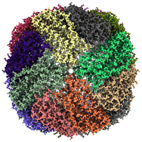
In cryoEM, frozen-hydrated samples are imaged using phase contrast without any chemical contrasting agents. The aim of cryoEM is to obtain a structure of a specimen as it exists in vitro, e.g. in a test tube, or in vivo, e.g. a living cell. Understanding the structure of a specimen is key to being able to understand its function. Typically, specimens for cryoEM are flash-frozen using a plunger- or guillotine-like device where they are immersed in a cryogen, such as liquid ethane, liquid propane or a mixture of the two at rates of over 1 m/sec. Essential thinning of the buffer across the grid just prior to flash-freezing is accomplished using blot papers. Alternatives that omit the blotting step have recently come to market. These alternatives also aim at coping with a critical component of the process, which is the air-water interface. The amorphous nature of the water of the cryo specimens requires the grids to be kept below -160ºC at all times, requiring the TEMs to have special remediations to prevent frost from building up on the samples. Every JEOL cryo-TEM has a special designed cryo box to accomplish this crucial task. Thus, samples can be kept frost-free for multiple days. Imaging cryoEM samples is done through low-dose techniques using sophisticated movie-type detectors from Gatan or Direct Electron followed by extensive image processing schemes, such as those in cryoSPARC or Relion. Although cryo-EM has been around since the 1980s, only recently (past 5 years or so) have enormous strides been made understanding issues like beam-induced motion, doming of the ice layer, and charging that have hampered the progress in this field for so many years. The reward of effectively dealing with these issues is obtaining structures of protein complexes at resolutions that allow pertinent questions about function to be raised and answered. The figure shows the 1.29Å structure of mouse apo-ferritin obtained by Fujita et al. on the
CRYO ARM 300 at Spring8 (Japan).
The bottom line: for every Life Science Application, there is a JEOL TEM perfectly suited for the job!
JEOL SEMs provide high resolution montages and three-dimensional images of structural details. JEOL FE-SEMs enhance imaging and throughput capabilities with image quality and resolution that rivals a TEM. An ideal tool for research and teaching facilities, the FE-SEM can be equipped with a Transmission Electron Detector, 3View Tomography and other analytical accessories.
JEOL FE-SEMs enhance imaging and throughput capabilities with image quality and resolution that rivals a TEM. An ideal tool for research and teaching facilities, the FE-SEM can be equipped with a Transmission Electron Detector, 3View Tomography and other analytical accessories.
JEOL Mass Spectrometers are used for protein characterization, proteomics, pharmaceuticals, drug development, and high throughput screening.
JEOL NMR spectrometers are used in a broad range of applications, from analysis of molecular structure, dynamics and interactions between various molecules, to fundamental material studies. Those applications are extensively used in many fields and areas, including drug research and development, structural molecular biology, development of battery technologies, food analysis and more.