April 26, 2007 (Peabody, Mass.) -- JEOL USA announced today that researchers at Harvard University’s Department of Molecular and Cellular Biology have selected JEOL as a partner in a collaborative effort to map the brain using high resolution SEM images. Harvard biologist Professor Jeff Lichtman, post-doctorate Narayanan (Bobby) Kasthuri, and University of Southern California Research Assistant Kenneth Hayworth plan to use a JEOL scanning electron microscope (SEM) to ultimately produce a 3D image of the entire mouse brain. Harvard will take delivery of a JEOL high resolution field emission SEM in June 2007.
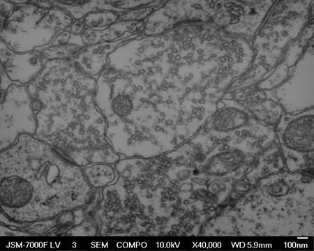
Working together, the JEOL and Harvard teams will customize the JSM-7001FLV, one of JEOL’s most popular SEMs used for advanced imaging and research, to sequentially allow fast acquisition of hundreds of thousands of high pixel density images at the nanometer scale. “We’ll ultimately be using image recognition software for montaging at very high pixel resolution – approximately five nanometers – to acquire up to twenty megabyte images in about three seconds,” said Charles Nielsen, JEOL USA SEM Product Manager and Vice President.
“We’ve learned a lot about SEM and robotics,” said Kasthuri, who said the Harvard lab team chose to partner with JEOL both for the SEM’s capabilities and the help of the JEOL team. “JEOL is as excited as we are.”
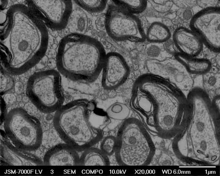
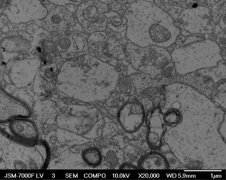
To produce the sample images, the Lichtman lab at Harvard is developing a new device that will automatically slice the sample and embed the organelles into a resin for sequential imaging. Hayworth, Kasthuri and Lichtman will head development and utilization of the Automatic Tape-Collecting Lathe-Ultramicrotome (ATLUM) designed to produce ultra thin (50nm and less) slices of the brain, and retain them in sequential order for reconstruction mapping. The entire sample, typically measuring 1mm x 1mm, will be embedded into a half-mile long substrate.
“We hope to eventually do cutting and data collection without human intervention,” said Kasthuri. “Not that it couldn’t have been done thirty years ago, but we couldn’t accumulate such large amounts of data before there was digital imaging.”
The Lichtman lab has used revolutionary techniques to study the brain, including sensitive laser and confocal microscopes to produce the “Brainbow,” which allows them to observe competitive interactions between neurons, providing insights as to how the brain is “wired.”
Lichtman’s laboratory specializes in visualizing synaptic rearrangements. The complexity of the 3D SEM imaging project has wide reaching implications that “may change biology a lot,” says Kasthuri, a Rhodes Scholar from Washington University with a Howard Hughes Medical Scholarship. Kasthuri has worked with Professor Lichtman to visualize changes at the junctions between developing nerve and muscle cells in mice. It is these changes in synaptic activity, and the selection process of synapse elimination, rearrangement, and competition, that most interests him.
“I am interested in studying the aging of the brain and how it matures,” Kasthuri says. “Why do we start life being capable of doing many things, and become very good at only a few things as we age? For example, why is it harder to learn languages as we get older?”
He sees this project as “adapting technology to arm scientists with tools that they need to make further inroads.” By creating a “wiring diagram” of the brain, Kasthuri says researchers may be able to learn what can go wrong and how to fix it when it does. “I’m also thinking really far out as to how we might influence things we don’t even know about yet, such as a computer that can see and robots that can walk as well as humans.”
The work will be presented at Microscopy & Microanalysis, Fort Lauderdale, Florida, on Tuesday, August 7, 2007, at 9:15 a.m. during the symposium “Multiscale Imaging of the Nervous System.”
Visit the Image Gallery on the JEOL USA website to see a Quicktime movie file of 30 sequential images of 50nm slices of mouse brain tissue imaged with the JEOL JSM-7000F. Serial sectioning done with the ATLUM Automatic Tape-Collecting Lathe-Ultramicrotome. This video is posted at www.jeolusa.com/tabid/472/Default.aspx.