 Peabody, Mass., June 26, 2006 -- JEOL USA has installed two transmission electron microscopes (TEMs) at the University of Texas Medical Branch’s (UTMB) newly-opened Cryo-electron Microscopy Center for Macromolecular Systems in Galveston, Texas. This facility contains the W. M. Keck Center for Virus Imaging, the first biosafety level 3 (BSL3) laboratory to house a cryo-electron microscope for high-resolution studies of deadly viruses and human pathogens.
These research tools will enhance UTMB’s frontline research capabilities in biodefense and emerging infectious diseases. UTMB is currently home to the National Institute of Health’s Center for Biodefense and Emerging Infectious Diseases and the site of the NIH-funded Galveston National Laboratory, the nation’s first National Biocontainment Laboratory.
TEM Imaging Used to Safely Study Live Viruses
Concerns about bioterrorism since the terrorist attacks of 9/11 have spurred additional investigation of viruses that could be, or have already been, exploited as biological weapons. The new cryo-electron microscopy (cryoEM) center will allow scientists to safely study West Nile, Venezuelan encephalitis, Rift Valley fever, hemorrhagic fever, dengue, SARS, avian influenza, and hepatitis C viruses, among other deadly pathogens. Renowned scientists at UTMB with extensive backgrounds in emerging viral infections will head the research programs carried out in the cryoEM center. UTMB will also use the facility to train young virologists for productive careers in developing new vaccines and antimicrobial therapies.
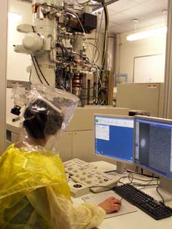
To study the virus samples, and to construct three-dimensional models to aid their investigations, researchers will use the JEOL TEM models JEM-2200FS 200kV field emission and JEM-2100 LaB6 cryo-electron microscopes. The first instrument is located within a BSL3 laboratory, and only accessed through three levels of security and safety barriers consisting of a series of rooms with door interlocks and decreasing pressure.
Biocontainment Enhanced by Remote Operation of TEM - Enables Scientific Studies Off-site
For additional safety and security, only researchers with appropriate government security clearance and advanced training are allowed in the high biocontainment areas. They will plunge-freeze the virus sample (inside a specially-designed glove box), load it into the microscope, and then leave the containment room to operate the microscope from a separate room through a network connection allowing real-time control of the instrument and all of its imaging capabilities, including the electron energy filter. These remote imaging capabilities will enable scientists working half way around the world to also access the TEM to conduct their studies without the need to be on site. With the necessary controls and Internet connection, they can remotely operate the cryoEM instruments from their own labs that are otherwise not equipped for this biosafety level of research.
“There are not many places to safely study these viruses,” said Dr. Stan Watowich, Associate Professor, Dept. of Biochemistry and Molecular Biology. “Galveston is a key center for virology research in the country. Any investigator wishing to work on highly pathogenic microbes such as SARS or avian influenza, and who has training and expertise in cryo-microscopy, can now do this work remotely at our facility.”
Watowich was part of the planning and development team for the new facility. He completed his post-doctoral work in structural virology with the late Dr. Don Wiley at Harvard, before moving to UTMB Galveston ten years ago. The facility is managed by Dr. Michael Sherman, faculty in UTMB’s Sealy Center for Structural Biology, whose major research interest is high-resolution structure determination of biological macromolecules and their assemblies by means of cryo-electron microscopy. These scientists were joined by Dr. Scott Weaver (professor in the Department of Pathology, and an international expert on encephalitic viruses), Dr. Wah Chiu (professor in Biochemistry and Molecular Biology at nearby Baylor College of Medicine in Houston, and one of the world’s leaders in cryoEM technology), and Dr. Henry Epstein (UTMB Chair of Neuroscience and Cell Biology, and renowned for his research on macromolecular assemblies) to develop, design, and deliver this unique facility.
JEOL Solutions for a Challenging Environment
More than two years of planning and construction led to the opening of the Center. In addition to creating a building designed for safe handling of these deadly pathogens, the planning team had to ensure that the microscopes they selected would be supported by the manufacturer in this challenging environment.
“We sat down with the JEOL team early in the design process to discuss different aspects of maintenance of the microscope and were impressed and quite pleased with their response,” said Watowich. “This should be the safest scope an engineer could work on. It will be certifiably clean.” In addition to strict precautions for maintenance and service, the JEOL team, working with Integrated Dynamics Engineering, Inc., of Woburn, Mass., provided a solution for isolating the TEM from electromagnetic fields and vibrations due to nearby elevators and the negative pressure air handling system. “The environment is less than ideal for these sensitive microscopes, but they are now performing at their optimum specification levels,” said Watowich.
“I believe that our experience with installations in less-than-ideal environments is a major plus for our service team,” said Patrick McGinley, General Manager of JEOL USA’s service division. “The JEOL team has worked very hard to investigate and test methods for sterilizing the microscope. We’ve also integrated with third-party providers to help determine the best means of thoroughly de-contaminating the instrument prior to routine maintenance or service.”