JEOL FE-SEM – Innovative Design
SMART – POWERFUL – FLEXIBLE
JEOL’s in column Upper Electron Detector (Through The Lens Detector) provides not only ultra-high resolution imaging but also includes a user selectable energy filter allowing the user to study a sample under different contrast mechanisms. For example, this energy filter allows the user to select low energy secondary electrons (SE) to enhance topographic features or high energy backscatter electrons (BSE) to highlight atomic number contrast. This detector is especially useful at lower kVs.
Mesoporous silica with Pd nanoparticles

UED with the filter set to SE (left); UED with the filter set to BSE (right)
The energy filter provides the user the ability to capture both secondary electron images showing individual particle dimensions, pore size, pore distribution and pore wall thickness and backscatter electron images showing the size and distribution of the active Pd catalyst particles with the same detector under identical SEM operating conditions improving ease of use. The in- the- column geometry of this detector yields high S/N ratio images for both BSE and SE Imaging.
Additionally the SEM can be equipped with a second in the column (through the lens) detector dedicated to SE signal collection (USD). This detector allows live signal collection (and live mixing) of both in-column detectors simultaneously creating images with both topographic and atomic number contrast. The energy filter can be applied over a wide range of acceleration voltages increasing its usefulness on a wide variety of sample types.
Au nanoparticles on TiO2
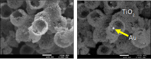
Surface morphology image using the USD (left); Composition image using the UED (BSE) (right)
The UED detector with the energy filter set to detect BSEs is ideal for generating images with atomic number contrast where the sample of interest contains thin layers on the surface. Using higher kV and traditional BSE detectors the beam penetration is simply too large and no contarst is observed. The UED with the filter allows very low voltage (sub 1 kV) imaging with strong Z conrast with a great signal to noise ratio. The examples below show contamination on a Li ionBattery electrode.
Li ion battery electrode UED BSE images at various kVs
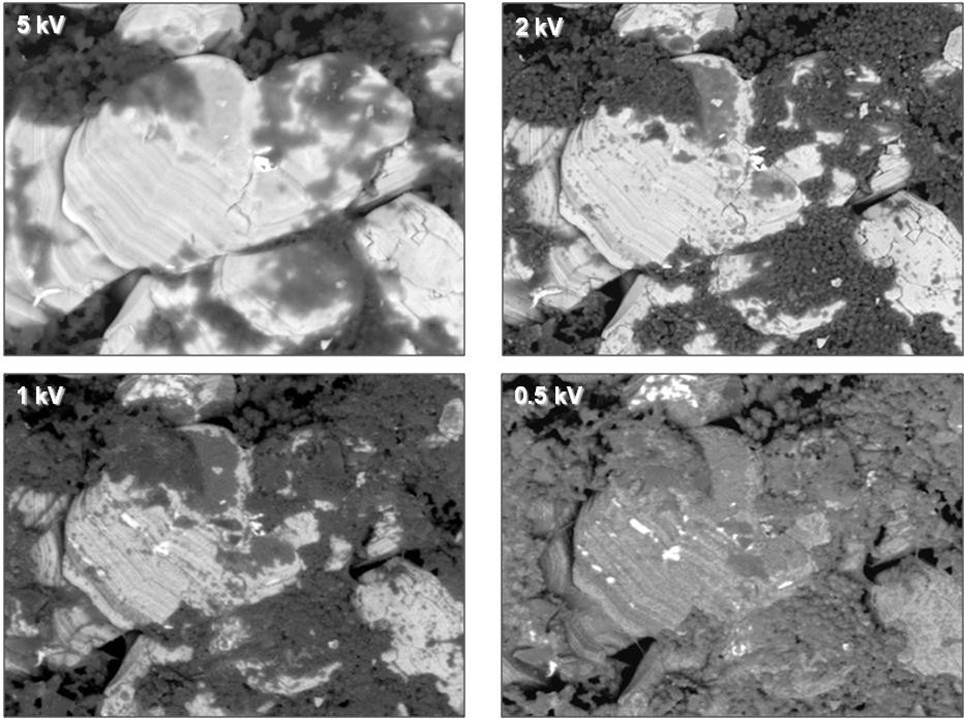
On rough samples at the low voltages required to see surface information the UED with the energy filter is more efficient than a traditional below the lens BSE detector for imaging Z contrast. The specimen topography( which is very useful information as well) is seen in the low angle BSE signal and the Z contrast is seen in the high angle BSE signal thus providing more information form the same area on the sample.
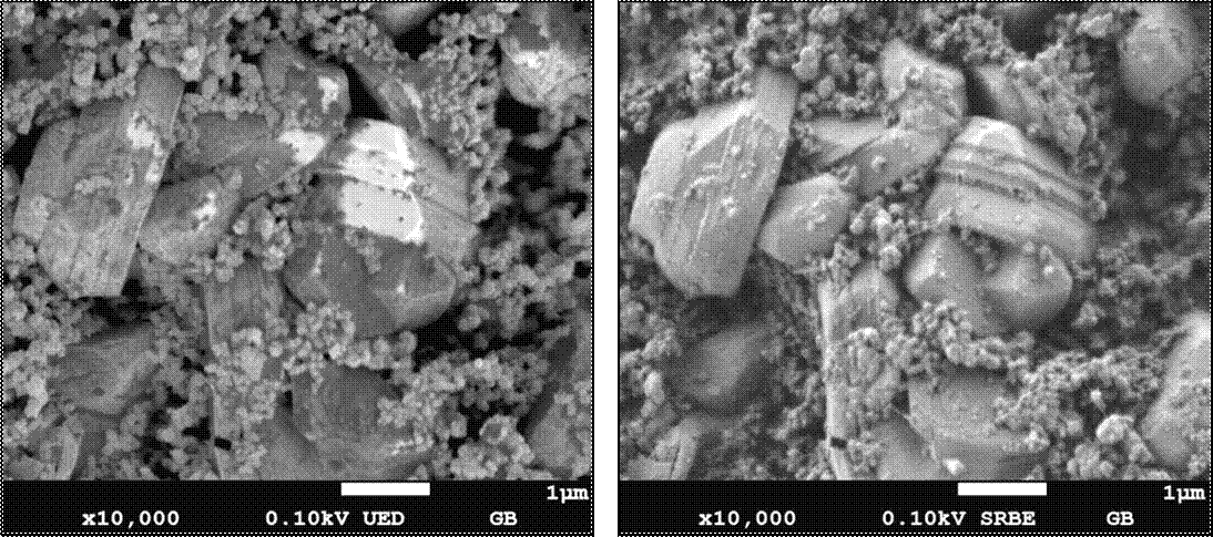
100V BSE UED image (left); 100V in chamber BSE image (right)