Microscopy in the Classroom: Bergen County Academies
There's no question that bringing microscopy into the K-12 classroom is helping to shape a new generation of scientists. However, schools that integrate microscopy into their science programs typically only have limited access to university instruments or use old equipment passed down to them. One public high school, however, has established its own lab with current instrumentation, and is taking an active role in the
Microscopy Society of America (MSA) Education Committee to help other educators advocate for and enhance their science programs.
Unique Educational Experience - Nano-Structural Imaging Lab
Bergen County Academies (BCA), a public "magnet" high school in Hackensack, New Jersey, established a Nano-Structural Imaging Lab (NSIL) in 2009, and equipped it with imaging instrumentation including a
JEM-2100 LaB6 TEM with cryo capabilities.
At BCA, more than 120 students to date have used the TEM for research projects. Some of these projects have been presented as posters at the annual M&M conference, further giving these students real world experience in their scientific endeavors.
The collaborative facility is accessible to students from any of the seven academies at Bergen County and interdisciplinary research is encouraged, but most are enrolled in the Science, Medical and Engineering Academies, working in cell biology, chemistry, nanotechnology, and stem cell research programs. They learn to use the TEM and other equipment, then how to apply it to their topic of interest.
"The research program at BCA is a unique educational experience for students that take part in it, showing them how to be a research scientist by developing and carrying out an independent research project based on the primary literature, in other words, learn science by doing science," Craig Queenan and Alyssa Calabro, instructors, and Dave Becker, Lab Manager explained.
In the NSIL, "The TEM is used for analysis and verification of nanoparticles, many of which are synthesized in the lab. It's also used in biological studies, where visualization of internal modifications to cells and tissue after treatments is necessary. Without powerful tools like these, students would not be able to perform the full analysis needed on their samples and provide the high quality image data to support their findings."
Students Eager and Engaged in Front of the Instruments
Although the TEM may seem more imposing than an SEM, students learn it "as quickly as they would a new video game or computer program. They were raised in an age where technology was not feared, and they are eager and engaged in front of the instruments...after the one to two one-hour training sessions on the TEM that we provide to acclimate them to the system and operation, the students begin to be as comfortable with the TEM as they would a digital camera." Quick start guides created by Queenan and Calabro provide step-by-step directions students can follow to turn on and saturate the filament, do sample alignments, image, and shut down after use.
Both Queenan and Calabro are Certified Electron Microscopy Technologists through the MSA. Queenan has a BA in Genetics and History and an MS in Molecular Biology from Montclair State University, and Calabro has a BS in Biochemistry and is pursuing an MS in Molecular Biology from Montclair State University. Becker began his career at the Academies 20 years ago and, with responsibility for new program development, has seen the school's transition from traditional vocational education to the unique learning environment that it is now.
Use of the TEM increases every year at BCA, and at M&M 2012, the microscopy industry's largest conference in the U.S., four scientific posters were student projects. This hands-on microscopy experience is accessible to students conducting research in the school programs, through collaborative projects with outside institutions, or through internship opportunities for students from other district programs.
MSA Outreach Subcommittee
The Microscopy Society of America's Education Outreach Subcommittee has made great efforts to promote microscopy in the classroom. BCA's Queenan, Calabro, and Becker held a special conference education event at M&M entitled "X-90: Microscopy in the Classroom Strategies for Education and Outreach." Some of the committee's additional efforts in K-20 education include:
- Project Micro provides information on using microscopy in the K-8 setting
- Family Affair and Microscopic Explorations provides hands-on activities for attendees and kids encouraging them to explore the microscopic world
- HS_SEM Google Group links programs around the world and offers a forum for discussion
Engaging the high school student in scientific research, and actually establishing a lab, was a feather in the cap for this unique public school. Through funding support from the Bergen County Board of Chosen Freeholders and Federal Perkins Grants, this New Jersey high school is making a difference both for students in the program and for local industry, universities, and medical facilities who benefit from the collaboration.
"Sparking creativity in the high school classroom will lead to a new generation of accomplished, motivated American scientists," Queenan said.
Joshua Meier is a Junior at BCA with an incredible background and many interests. He explained his research using the TEM.
What is your topic of research?
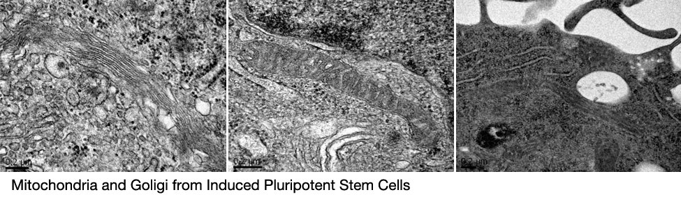
Induced Pluripotent Stem Cells (iPSCs) have extreme clinical potential, can be artificially generated in the lab, and can be tailored for an individual patient. Unfortunately, these cells are victim to several problems that cause them to die prematurely and they are therefore not currently used by physicians. When I first started my work at the Academies, I wondered why these cells have problems compared to classical embryonic stem cells, which are medically used. Through my research, I investigated novel properties of these lab-engineered stem cells and found that they are victim to deletions in their mitochondrial DNA -- the missing genome that has largely not been considered by many scientists. In fact, I found that over 1/3 of the entire genome disappears when these cells are created, finally shedding light on the reasons why these cells have problems and pointing to explanations of how these stem cells actually work. I have now turned my investigation in a new direction and am applying my findings to another field, cancer research, in order to use my findings to develop new strategies toward fighting the disease and learn more about both stem cells and cancer.
How has microscopy been used in this project and how has it helped you?
After discovering that these stem cells have major deletions, I wondered whether these profound genomic changes would have affects on other parts of the cell. Using transmission electron microscopy, I analyzed the stem cells and compared them to the cells they were made from. I found that the mitochondria of the stem cells underwent a radical transformation -- they became elongated and huge.
Two years ago, as a freshman at the Academies, I heard about a class that was offered called "Research Applications to Molecular Biology and Genetics". I had taken university-level classes in genetics the previous summer and I simply took the class to broaden my knowledge. Little did I know that one of our assignments - a research proposal - would later turn into my own real project.
When I first began my research, I had never considered the potential applications that microscopy could have. However, after my results demonstrated the extent of the deletion I was studying, I began thinking of ways to determine whether these deletions actually made a difference to the cell. Later, when I was sitting in my nanotechnology class, the idea struck me. We were learning about electron microscopy and I pondered whether I could incorporate the technology into my project. I found the answer and made another extension to my project. I started working in the nano-scale imaging lab immediately, and gained valuable experience in a new field.