Children's Hospital Colorado
The electron microscopy lab at Children's Hospital Colorado never loses sight of its underlying philosophy: Children not only are their ultimate customers... but, as its name indicates, are also the true owners of the institution that employs them. Dr Gary Mierau emphasizes that the rewards for providing this service can take many forms… the most gratifying of which is when a former patient, writing a school report about the time he/she had cancer, returns to see the equipment, the people, and the tissue specimen involved in establishing their diagnosis. This keeps it real!
The laboratory has been in existence since 1968. The Scientific Director and four full-time electron microscopists have more than 100 years of combined experience in the field of diagnostic ultrastructural pathology. The staff have authored more than a hundred scientific articles, reviews, book chapters and books on ultrastructural pathology and lectured in more than a dozen countries. It is a high volume facility dedicated to delivering an exceptional product with extraordinary speed (normally within 24 hours of specimen receipt). The laboratory is equipped with two modern transmission electron microscopes (one being a JEOL JEM-1400Plus) and a scanning electron microscope (JEOL JSM-6010LA). By maintaining a long term vision and careful succession planning in the form of equipment purchases as well as the hiring and training of quality electron microscopists, the EM Lab at Children’s Hospital Colorado is poised to remain an important diagnostic lab for the foreseeable future.
In addition to conventional electron microscopy, specialized diagnostic techniques such as energy dispersive x-ray spectroscopy for heavy metal detection, tomography for 3D reconstructions, scanning electron microscopy for hair shaft abnormalities, direct negative staining for virus identification, and whole-mount studies for platelet granule quantitation are offered.
 TitleCategory
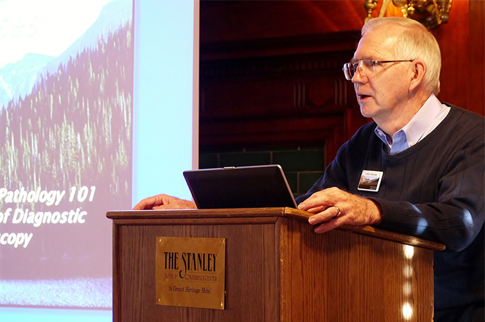
Dr. Gary Mierau speaking at the introduction of the Ultrapathology 101 course
 TitleCategory
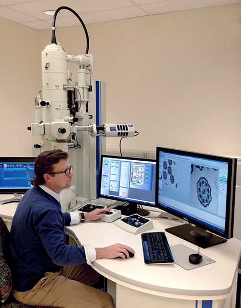
Eric Wartchow at the JEOL JEM-1400 Transmission Electron Microscope
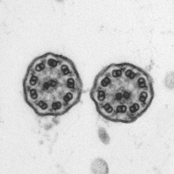
The unit provides local, regional and national clients with a full range of diagnostic services. The staff has extensive experience with renal, respiratory, muscular, metabolic, hepatocytic, neoplastic, infectious, hematologic, and other disease processes. An area of particular interest and expertise is with the diagnosis of diseases involving the respiratory cilia. These present a complex set of challenges with regard to specimen processing, imaging and interpretation… the specimens are small and dispersed, the structures of interest fall at the limits of obtainable resolution and the diagnostic criteria are imprecise. As a consequence, such specimens often are referred even from institutions operating their own diagnostic facilities.
Eric Wartchow, who is nearing completion of his PhD studies, has been leading the team in this area for the past ten years. He has authored several papers on the topic, including one identifying a new disease variant. He currently is engaged in an assessment of the diagnostic utility of 3D tomography (photo) with the hope of establishing a superior means of visualizing the critical internal structures of the cilium when dealing with suboptimal specimens.
 TitleCategory
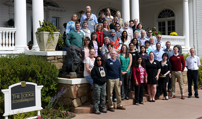
Ultrapathology 101 course attendees at The Stanley Hotel, Estes Park, Colorado, September 2015
 TitleCategory
An area of particular interest and expertise is with the diagnosis of diseases involving the respiratory cilia
Defying conventional wisdom, the unit has demonstrated that it actually is possible for a diagnostic electron microscopy laboratory to be a revenue center, rather than a cost center, within its institution. This is largely made possible by a significant referral business. Three-fourths of the diagnostic caseload is currently being received from external clients. While purposefully not functioning as a “core lab”, a modest secondary revenue stream flows from the provision of “spare-time” assistance to researchers not able to obtain the needed assistance elsewhere.
Because the institutional mission identifies education, research and advocacy (in addition to patient care) as being essential to improving the health of children, the unit is also purposefully engaged in addressing each of these elements of the mission. In addition to providing a structured component of the resident/fellow training program, last autumn the team organized and presented an intensive four-day course, which drew attendees from around the world, entitled, “Ultrastructural Pathology 101: Fundamentals of Diagnostic Electron Microscopy.” The team also contributed a number of book chapters and research publications, made platform and poster presentations, provided peer reviews, and served as executives in the Society for Ultrastructural Pathology. A lot of activity… a lot of fun!