Lichen Labs
"Looking at nature from a materials scientist’s standpoint is very different from how a biologist looks at it," Sue Okerstrom says. After 25-years of looking at polymers, metals, and ceramics at Medtronic, she retired early to pursue her passion for biomimicry - innovation inspired by nature. Sue established Lichen Labs to help industry, architects, and students explore how nature can guide us to innovative new materials, processes, structures and textures to solve human challenges on all scales.
Biomimetic products already on the market include the Japanese bullet trains based on the structure of the splashless Kingfisher beak and head to stop sonic booms when going through a tunnel, chainsaw blades using the same shape and cutting action as woodborer beetle larvae to cut trees down more efficiently than axes, and in the medical industry, sharkskin-inspired surface texture that repels bacteria with no chemicals or antibiotics.
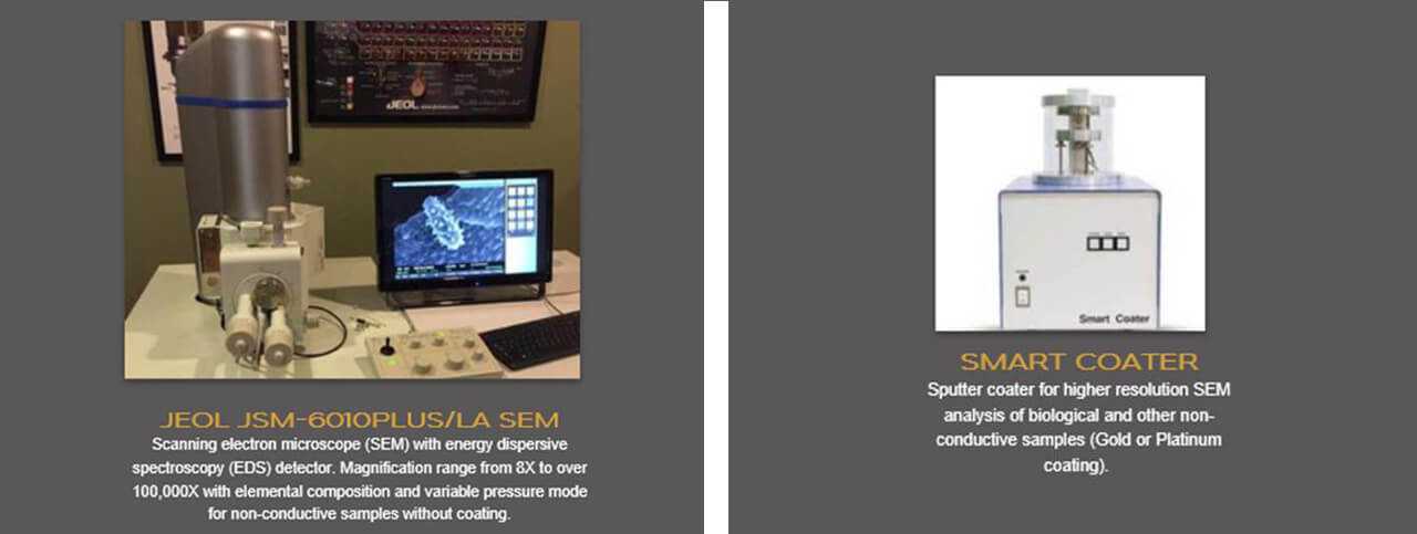
After a few structural changes to her home in Minnesota, Sue installed a JEOL electron microscope, a stereo microscope, compound microscope, built a 3D printer, and she was on her way to providing imaging and analytical services plus a Macro to Micro education program and biomimetic research.
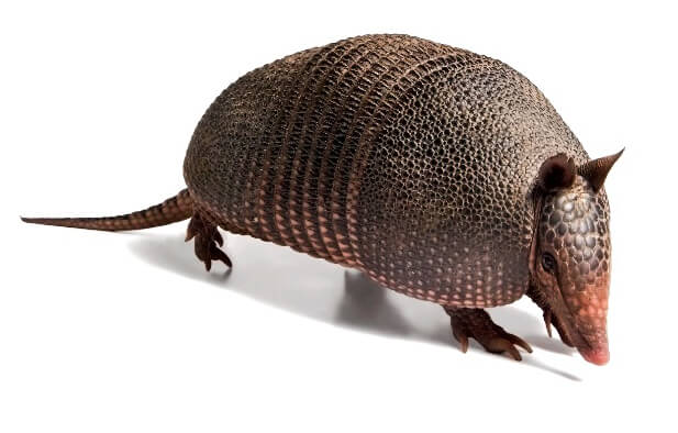
You might say it all started with an armadillo. The mammal, with shields of hard, protective scales sparked her imagination during an innovation conference that included biomimicry. Sue thought in that moment “This is what I want to do with my life.” She found a certification program, “Biomimicry 3.8”, a title implying that life has been on earth for 3.8 Billion years and has much to teach us today. As one of a 20-person cohort she pursued a professional certificate that was accredited during the program as the first MS in Biomimicry from Arizona State University. It was a two-year adventure of a lifetime, traveling to six unique ecoregions in the world and immersing herself in the environment and ecology. From Botswana to the Sonoran Desert, Vancouver Island, Costa Rica, and the Blue Ridge Mountains of North Carolina to the mountains in Montana, each region opened her eyes to how organisms adapt to their environment. She saw firsthand the possibilities for new designs and manufacturing techniques that nature could teach us.
The class members were chosen for the diversity of their backgrounds - biologists, engineers, designers and business specialists - so that they could each bring something different to the table from understanding the biology to the design and marketing of new products and materials. Then, they would work on the projects together or individually with their own expertise. Every member played a vital role. For Sue, she added materials and microscopy. The time spent working together in the cohort developed and strengthened connections and formed collaborative partnerships that are still maintained today.
“It was very hard to go back to normal life,” she marvels, yet she has brought her rich experience back to create Lichen Labs LLC. The name of her company was inspired by the orange lichen she found in Montana. As for the company logo, she explains, “My mother carefully carved off a similar orange lichen, Xanthoria elegans, with her pocketknife from a tombstone in Virginia MN.” The JEOL 6480LA-Plus SEM was used to successfully image the large sample at low magnification with high resolution in variable pressure mode, ultimately becoming the model for the Lichen Labs logo. Note the 0.5mm (500um) bar.
The width of the lichen shown on the right is 6.65 mm.
Lichen are a marvel of nature. They exist without roots, covering around eight percent of the terrestrial surface of Earth. A fascinating attribute of the organism is its ability to completely dry out (sometimes for decades) and come back to life to begin photosynthesis as soon as moisture is returned. Some types of lichen can live for over 4000 years! She examined the Montana lichen with the electron microscope and explains, “I did a BEC image and noticed bright areas around the rim of the cup-like structures.
“So, I did EDS and it was almost pure tin. A very unexpected result. The tin was collected from the air through the pores of the lichen and concentrated in the rim of the cup-like structure.”
Case Study – Orange Lichen.
“I found out later that Lichen are miners. They are analyzed in Australia to indicate where gold could be mined. I also didn’t realize lichen were not a single plant organism. Fungus is the body and algae and or cyanobacteria lives in the body and provides food through photosynthesis. Lichen are a symbiotic relationship – like the collaborative nature of Lichen labs - “symbiotic partnerships for scientific solutions”.
To further her efforts of encompassing biomimicry in everyday products, she has ventured into applying microscopy to biomimetics, both as a discovery and a problem-solving approach where one seeks to understand and emulate aspects of biological adaptation in applications in the human world. With a classmate who is an architect, they are looking at developing stronger building materials, drawing inspiration from
tree nut shells that don’t easily degrade due to their high mineral content.
You can hear her sense of wonder as she describes some of the valuable lessons she has learned from nature. Her enthusiasm stems from her own relationship with the environment and her ability to share her knowledge with others. Whether it's watching the 7th graders’ faces light up as they design their own nature-inspired creations in Life Science class, or simply exploring the beauty of the physical world during one of her kayaking, hiking or skiing adventures, Sue carries a deep appreciation for the wisdom Mother Nature provides wherever she goes.
In addition to working with industry and researchers in different fields, Sue has developed a “Macro to Micro” STEAM (science, technology, engineering and math with an “A” for art) pilot program aimed at including biomimetic education and outreach utilizing microscopy in middle school curricula. The program is in the scale up process to have Universities do the outreach to 7th grade Life Science classes. The University of Minnesota is currently supplying a graduate student for the 2019-2020 school year and donating SEM instrument time. Outreach is to a handful of schools with trained teachers in Ely, Minnesota, the flathead reservation in Montana, and the Boston area.
Macro to Micro STEAM Program
The students look for a specimen in nature such as a feather, plant part, or insect that can be examined in an electron microscope. They identify things they are interested in studying and research structural adaptations at the micro and nano scale that can be mimicked. Using their creativity, students will then invent their own product based on the function of their specimen’s structural adaptation that could be used in the human world. The teacher sends in 6-8 highlight samples to be analyzed at Lichen Labs or a University Lab by a graduate student. About a week later the teacher remotes into the microscopes in the lab hundreds or thousands of miles away and can actually run the microscopes and see their samples from macro to micro to help them better understand the concept of scale and maintain context.
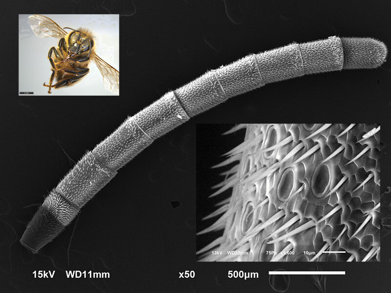
Above is an example of what students might see on a specimen. The top left picture is a honeybee in the stereo light microscope magnified 7X. An antenna from the bee is put into the electron microscope magnified 50X in the center. We can see that the antennae has different electrical charges. The dark gray area at the base is more negatively charged and the tip is slightly negatively charged compared to the rest of the antennae. To better observe the differences, the antenna is magnified 1600X in the inset picture. So what’s going on? It turns out that the ground is positively charged, and flowers are negatively charged. When the bee flies through the air it becomes positively charged by the air particles hitting it. As it starts to land the negative pollen is attracted to it and it changes the field on the flower to be more positive. This field change signals other bees not to stop at that flower since it has already been visited and the nectar supply is likely low.
In the picture below, the antenna surface is magnified further to 3500X below showing the satellite disc like structures and sensory hairs. These features give bees the ability to sense the Earth’s magnetic fields, enabling them to travel further distances and detect flower scents at a closer range. Bees already play a significant part in our ecosystem and benefit a plethora of species including humans. With their incredible anatomy, the bee could go as far as inspiring a new type of electromagnetic field navigation system based on their antennae.
Learning from nature – whether it’s studying structural color in moth and butterfly scales (below) or the superhydrophobic skin and air trapping structures on her living “Fishing Spider”, Sue is opening new horizons.
She developed a method to image the cross-section of a single Lepidoptera scale in order to understand more about differences in structural interaction with light between black and white scales in moths and butterflies in collaboration with the University of Minnesota. These studies may inspire new materials for thermal regulation in buildings or clothing.
Above a 3 ½” diameter Dolomedes scriptus fishing spider was found perched on a nursery web at the Manitou River in Northern Minnesota. This one was unexpected and keyed out later via photographs. “At the time I had not heard of a fishing spider. Once informed I caught a Dolomedes tenebrous in another location. The Dolomedes are opportunistic predators that preys on small aquatic or terrestrial invertebrates and vertebrates. It hunts for prey near, over, on and in water rather than capturing it in a web. It can walk on water and discovers prey such as small fish through disruptions of surface tension of the water” she marveled.
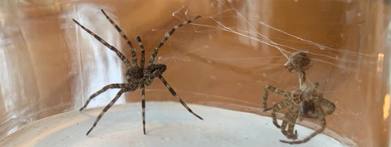
“Charlie” shown with molt complete with fangs at right. In order to get larger, Charlie grew a whole new spider inside this exoskeleton that was connected by a membrane. When it was time to molt an enzyme was exuded dissolving the membrane away. Charlie increased her blood pressure and literally popped off the top of her head and pulled herself out with the aid of a web strand – like climbing out of a wetsuit. Once she molted, she was too large to be a “he”. Male and female differentiate by size.
Exoskeleton molt foot (left); Bush like hairs that trap air on molt (right).
Hairs were cut to expose hair follicles and superhydrophobic skin, magnified above right. Fishing spiders can breathe underwater (through its superhydrophobic skin and structures trapping a layer of air around its body). When it goes through water there’s almost no friction. Known as the Salvinia Effect, this ability is being studied for its drag-free properties and will inspire new marine products, perhaps.
“I see things so differently than before. All structures are built for particular function(s) and the mechanism for carrying out each function is efficient and streamlined,” she says.
Sue began her career in mining, with a degree in environmental science “back in the days when it was a new type of major and there were no jobs.” She launched her career at US Steel in Minnesota as a metallurgical technician working in process control at pelletizing plant for taconite. She went on to work in electrode and feedthrough development at Medtronic. While there Medtronic supported obtain a master’s in materials science and engineering. After successive promotions, Sue was named a Technical Fellow at Medtronic, served as President of Minnesota Microscopy Society, started Lichen Labs and will serve as Co-chair at M&M 2020 in Milwaukee for biomineralization and biomimetic materials.