Microtrace LLC
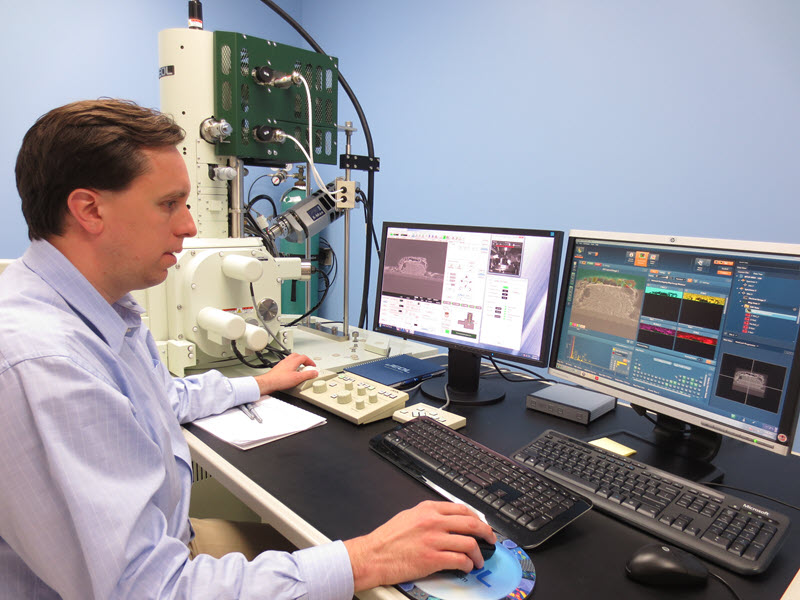
Given the high-profile forensics cases handled by Microtrace, the company’s father-son team of Skip and Chris Palenik have achieved a kind of celebrity status. But more than accolades, they relish the challenges sent their way and the opportunity to summon their vast expertise to let science determine the answers to the questions posed. These questions sometimes put them in the public eye. In the fall of 2018, the second season of the popular Netflix documentary, “Making a Murderer,” aired on television, showing Chris
using a JEOL Field Emission Scanning Electron Microscope. In episode 7, he examined key evidence that might help the defense’s case for a mistrial. Could the lead bullet fragment found in the garage of the defendant have passed through the skull of the victim, and were the particles embedded in the bullet fragment actually bone? After six months of waiting for a court order to release it specifically for this purpose, the lawyer watched as the embedded particles in the bullet fragment were carefully examined in situ at Microtrace’s lab in Elgin, Illinois.
 Microtrace LLCCategory
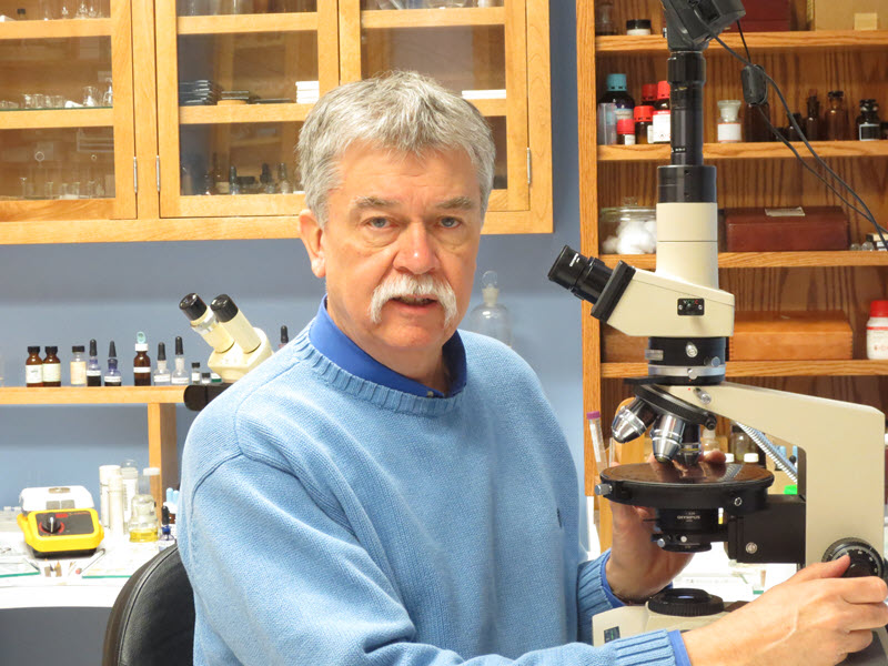 Microtrace LLCCategory
Spoiler alert! Using SEM imaging and Energy Dispersive X-ray Spectroscopy (EDS) analysis, Chris found the particles to be wood, not bone, supporting the defense’s position that this was not the bullet that had killed the victim. Chris says he isn’t really sure what the outcome will be in the case, he just gave his findings with the neutrality and careful consideration for which the lab is known. “We really work hard to provide answers that actually help somebody move an investigation along,” says Chris.
Creative problem solving through microscopy & microanalysis
What sets the Microtrace lab apart is the creative approach to problem solving through microanalysis. “Our work is based on solving a problem, whereas a lot of people that have labs work on processing huge volumes of samples in the same way. It’s part of the reason we’ve had success in forensic cases. While we are ISO-accredited, and sometimes have to use a standardized approach, we try to use creativity and a sample-driven approach to look at each sample and look at the question being asked to try to come up with best analytical path forward.”
In addition to having looked at evidence for several famous murder cases, their investigations have ranged from examining art, collectables, sports memorabilia, historical artifacts, geological samples, and industrial products. “We do a lot of work for a wide range of industries helping them solve their problems, whether it’s foreign materials in their products or helping them understand the design of new materials.” The collectables, art, and historical items are “always kind of fun to see because they’re unusual – whether it’s a painting that’s possibly 500 years old or a baseball bat that Joe DiMaggio was swinging, we get a lot of very interesting questions.”
Their diverse interests and a lifelong passion for investigating using microscopy began early on, when Chris’s father, Skip had his own microscopy lab in their basement. Skip worked closely with his mentor Dr. Walter McCrone, a renowned expert in the microscopy field. Scientists and investigators, including one from Scotland Yard, stayed at the Palenik’s home when Chris was a child. Like his father, Chris was introduced to the microscope very early in life. He attended classes at the McCrone Research Institute as a middle and high school student. Skip founded Microtrace in 1992 and the father and son have worked together ever since. The company has grown to 13 staff. Their knowledge spans many, many fields, and they keep a wide range of databases and physical reference collections to consult - over 35,000 curated specimens ranging from sand, soil, glass, hair, pigments, dyes, food ingredients and more. Chris has been invited to speak at the National Institute of Standards on curating databases.
A closer look with Field Emission Scanning Electron Microscopy
 Microtrace LLCCategory
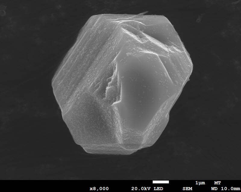
A crystal of zinc sulfide used in a fluorescent tracer that is used in police investigations
 Microtrace LLCCategory
Ion milled toner particle from research into forensic applications of nanoparticles and nanoscale features
 Microtrace LLCCategory
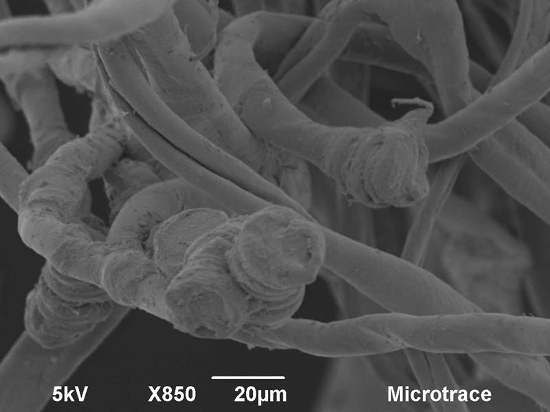
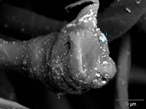
Polyester fibers severed and melted by an AK47
 Microtrace LLCCategory
BSE image of polyester fibers where you can see the lead particles
 Microtrace LLCCategory
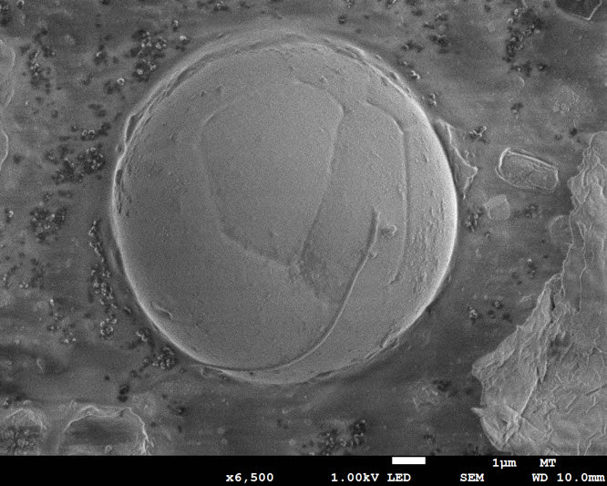
GSR particle observed at low voltage to see surface detail
Microtrace uses a Field Emission SEM as well as a tungsten SEM in their lab. The ultrahigh resolution FE SEM allows imaging at extreme magnifications. Most crime labs do not have this level of capability, because a tungsten SEM Is more affordable, and answers most of the “basic” questions in forensic science.
“The FE SEM allows us when necessary to look closer. Crime labs typically look at particles probably on the order of 10-100 microns on the low side with the exception of gunshot residue (GSR) which goes down to a half micron. There is not much exploitation of evidence that is smaller than 10 microns. We look at that quite commonly – just about any material you can imagine –in various cases we’ve applied SEM analysis to look at particles even smaller than one micron.” Consequently, Chris says, “we’re working on a grant on developing nanotrace evidence for forensic science and are looking at basic questions about scaling analyses down further.”
An ideal application for the FE SEM was to look for traces of lead particles in a bullet hole1, using a simulation of the event, to provide evidence used in a military court martial involving a claim that a hole in a cotton-polyester blend shirt was caused by a bullet. There was no available data on what constitutes a bullet hole in fabric, so the simulation was conducted, resulting in microscopic traces of lead particles embedded both on and in the melted fibers. While GSR had already been identified from a sample collected near the hole, a closer look determined that if a bullet had passed through the fibers, the synthetic fibers would have melted and snapped back on themselves and lead microparticles would be adhered to and embedded in them. SEM and EDS were used to locate and analyze particles of lead in the simulated sample. Ultimately it was determined that the hole had been made with scissors, not a gunshot.
In a child abuse case, the SEM was used to find microscopic traces of makeup in a discarded tissue, with barely any visual indication to the naked eye. Preliminary examination with a light microscope showed there were spheres of material they initially assumed was oil. In the SEM they determined that the spheres were actually silica. “The SEM allows us to tell if the spheres were solids and their distribution in situ. The material was both silicon and oxygen – seen without carbon coating - and several mica flakes. It doesn’t say for sure it’s makeup but, in combination with other components, it puts it in the realm of plausibility to form the conclusion that it was.” They are adding an Electron Backscatter Diffraction (EBSD) detector to their analytical capability with the SEM. “I’m very excited - I see a lot of interesting applications for forensic geology looking at particles with EBSD and the phase ID aspects of it.”
Paint sample preparation is key
For the ultimate in preparing paint samples for examining in the SEM, they use a JEOL ion beam Cross Section Polisher that produces pristine surfaces for imaging and analysis at high resolution. “Typically, in paint analysis the sample prep is quite poor to the point where it’s hard to resolve some of the thinner layers in the SEM. Regular microtomy can produce pretty good samples, but most labs use a scalpel under a dissecting microscope where they scrape off layers of paints. A lot of paints have sub 10 micron layers so it’s a very challenging thing to do freehand,” says Chris. The ion beam polisher makes an enormous contribution to paint sample prep.
Chris is part of a materials committee revising documentation for paint analysis. The lab is internationally-acclaimed in advancing the field of microanalysis, and both Skip and Chris have been invited speakers at major conferences, consulted on countless forensic topics, including counterfeiting, and also teach forensic-related classes at McCrone Research Institute.
Where in the world did it come from?
With degrees in Chemistry and Geology from University of Chicago, and an MS and PhD in Geology from University of Michigan, Chris has a special area of expertise in soil samples, and Skip has developed the analytical approaches to soil analysis used by most forensic science labs. The sample analysis is broken into different fractions, including the light mineral fraction and heavy mineral fraction. Polarized Light Microscopy (PLM), Raman and SEM EDS help identify the minerals in the soil. Chris conducted exploratory work looking at polished (and unpolished) grains with SEM, EBSD and EDS. In addition, markings on the surface reveal a lot about the environment from where the soil or sand came. A case they investigated involved a shipment of cell phones that had been stolen and replaced with sand. By breaking down the mineral composition of the sand and consulting their reference collections, they were able to narrow down the port of call where the theft occurred.
1 Palenik, C.S.; Palenik S.; Diaczuk, P. Plumbum Microraptus: Definitive Microscopic Indicators of a Bullet Hole in a Synthetic Fabric. THE MICROSCOPE. Vol. 61:2, pp 51–60 (2013)