Imitation of Life – Alphaviruses Inspire Metamaterials at Indiana University
The acquisition of a new 300 kV Transmission Electron Microscope (TEM) from JEOL distinguishes Indiana University, Bloomington, Indiana as a major United States research facility where scientists can examine both biological and materials science structures at nanoscale resolution.
In February 2007 an NSF Major Research Instrumentation Grant was awarded in parallel with a $1M investment made by the University’s College of Arts and Sciences. The state-of-the art TEM will be housed in the University’s newly-constructed multidisciplinary science building, Simon Hall.
The TEM, a model JEM-3200FS, will be used for multiple disciplines by students and researchers in the fields of biology, chemistry, materials science, biochemistry, environmental and evolutionary sciences, geology, microbiology, and behavioral science - reflecting the University’s stated philosophy of achieving new vistas of understanding through interdisciplinary research: “If we unite great scientists of diverse backgrounds, they can solve problems together that they cannot solve alone.” Biologist and Assistant Professor Tuli Mukhopadhyay serves as Principle Investigator on the five-member team responsible for acquiring the TEM that will meet the requirements of several departments at the University.
Cryo TEM and 3d Tomography Used to Study Alphaviruses Dr. Mukhopadhyay’s biology lab studies alphaviruses which are transmitted by mosquitoes to a variety of hosts, including mammals. The virus is responsible for symptoms of arthritis, muscle aches, joint pain, fever, and certain alphavirus species can cause fatal encephalitis. The alphavirus particles, or virions, have an external diameter of approximately 700 angstroms. Dr. Mukhopadhyay’s lab will use the microscope for cryo-electron microscopy and 3D image reconstructions on virus particles as well as for tomography studies on viral assembly intermediates. Studies of Sindbis virus are important because these particles are very closely related to studies of the more pathogenic Flaviviradae viruses responsible for yellow fever, West Nile, and hepatitis C.
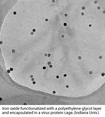
(images courtesy of Dr. Mukhopadhyay)
According to Dr. Mukhopadhyay, “Our primary tool for structure determination is cryo electron microscopy, which has become a powerful tool for determining three-dimensional structures of specimens that are too heterogeneous, too large, or too transient for conventional X-ray crystallographic methods. Rapid freezing of samples can trap viral intermediates and their real-time structures can be determined.”
The TEM will be used to produce an entire reconstruction of the virus particle using a combination of cryo tomography, 3d image reconstructions and atomic structures determined from NMR and X-ray crystallography. Researchers in the biology department will create a 3-D image reconstruction of the virion, and then begin to fill it with what they know about the proteins inside.
“We’ll fit in the atomic structure of individual proteins that make up a virus or the intermediates we have isolated,” says Mukhopadhyay. “It’s like putting together a puzzle. We have an idea of where different protein combinations would fit. It’s as though image reconstructions will give us the outline of the human body, while atomic structures of the proteins give us the thigh bone and the thumb bone and the shin bone. We’ll rely on biochemistry, genetics, and immunology data to figure out where everything belongs.”
Viruses Serve as Building Blocks for Metamaterials Reconstructing the viruses with the help of the TEM not only advances research in virology, it provides a model for nanotechnology in materials science.
Dr. Bogdan Dragnea, Assistant Professor of Chemistry and Adjunct Professor of Physics at IU, will use the TEM in his nanoparticle research. Dragnea’s research focuses on mimicking virus self-assembly in developing self-assembled molecular layers and nanoparticles for materials science applications. Dragnea’s group pioneered a technique to promote the self-assembly of a virus capsid around a functionalized nanoparticle.
Virus-like particles encapsulating a gold core have been also shown to have promising properties as building blocks for metamaterials. Metamaterials have optical (or more general, electromagnetic) properties determined by their organized structure rather than inherited directly from the material properties of individual subunits. The optical response of metallodielectrics is intensely studied at present because of their freedom of design and promise for novel properties, including better lenses, exotic coatings, new lasers, and miniaturization of photonic technologies beyond the diffraction limit. Research in Dragnea’s group has shown how biomimetic self-organization can be employed to combine the natural characteristics of virus capsids with the physical properties of inorganic nanoparticles -- the goal being to obtain metamaterials with novel optical properties.
Simultaneous Characterization of Inorganic and Biological Materials The key element in both research directions is understanding the role of the core-capsid interactions in assembly and structure of virus-like particle complexes. Here, both inorganic and biological material aspects have to be characterized at the same time, hence the requirement for a hybrid high-resolution/ cryo-em instrument. Lyudmila Bronstein, Senior Scientist at the Department of Chemistry, will design nanoparticles for enhanced magnetic resonance imaging and virus studies in collaboration with Bogdan Dragnea’s group. She has used TEM at high resolution to determine the fine structure of the magnetic nanoparticles (see fig 1), the properties of which are directly dependent of the nanoparticle size and structure. The advanced techniques of TEM sample scanning will allow mapping of different elements of nanoparticles thus revealing the composition of complex core-shell particles as shown in figs 2a and 2b.
Further Information
Tuli Mukhopadhyay's Faculty Page and Research Group.
Bogdan Dragnea's Research group.
Lyudmila Bronstein's Research Group.
Additional Research Planned for the TEM
Bacterial Morphongenesis and Cell Division Biological research of a different nature will be done to understand the basic mechanisms of bacterial cell division and cell differentiation, and to determine how and why bacteria undergo specific changes in morphology. Yves Brun uses Caulobacter crescentus and Hyphomonas neptumium as model systems to focus on the synthesis and function of the stalk appendage. Synchronized populations of swarmer cells, which have no stalks, can be isolated for both bacterial species, making these ideal systems for characterizing the morphogenesis of the stalk.
Brun’s group has taken a new approach and has begun to study the 3D structure of Caulobacter and Hyphomonas cells using cryo tomography. “Our goal is to obtain ultrastructural information of the morphogenesis of the stalk during the bacterial lifecycle,” Brun says. “We have already obtained mutants that are deficient in stalk synthesis or that synthesize stalks at the wrong subcellular site. We will analyze these mutants to determine the structural nature of their defects.”
Prokaryotic Cell-to-Cell Interactions Clay Fuqua’s laboratory studies the proteins and genetic networks involved in prokaryotic cellular interactions, specifically quorum sensing and biofilm formation. In particular, the lab has a strong interest in the structures and regulation of the components involved in these processes. laboratory works with Lingling Chen’s laboratory (IU Biology) to study the structure and association of TraR and TraM, two proteins involved in quorum sensing in the plant pathogen A. tumefaciens50. Atomic structures of TraR and TraM homodimers have been determined, but the proposed octomer complex has been recalcitrant to structural analysis, suggesting it may be heterogeneous or labile under crystallographic conditions. We need to use cryo-EM to determine the structure of the TraM/TraR complex. A three dimensional structure, determined by single particle reconstruction, can be obtained from a heterogenous population. The atomic structures of TraM and TraR will be placed into the cryo-EM reconstruction generating a quasi atomic structure. Quorum-sensing regulatory mechanisms. Adhesive surface structures within bacterial biofilms. The structures required for biofilm formation in A. tumefaciens are likely to be proteins, proteoglycans or polysaccharides54,55. “Our lab has attempted to compare wild type and mutant strains to identify polar structures on whole cells using fluorescence microscopy and TEM,” Fuqua says. “We have been unable to definitively visualize such structures, beyond the presence of simple flagella (fig. 13). Under cryo conditions, the delicate surface features will remain intact, and the FEG-TEM is ideal for visualizing and imaging the cells.
Juergen Schieber: Mudstones and the Hydrocarbon System The Schieber lab studies mudstones, fine grained sediments that constitute approximately two thirds of the sedimentary rock record. Mudstones are an important component of the hydrocarbon system because they are simultaneously the source rock of hydrocarbons, as well as the seal rock that later on prevents their escape from reservoirs. Because of their fine grain size, compacted and consolidated mudstones have very tiny pores, the size and geometry of which are critical parameters that determine whether in a given situation, a mudstone unit will be an effective seal for hydrocarbons, and also whether gaseous hydrocarbons that are adsorbed to the mudstone components can migrate out and support natural gas production.
Chen Zhu: Natural Nanoparticles and Mineral Surface Chemistry “Our lab quantitatively studies nanoparticles and mineral surface properties related to reaction kinetics, specifically the movement and distribution of mass and isotopes in groundwater systems due to fluid flow and water-rock-gas-microbe interactions,” Zhu says. The lab studies the sediment coating materials from the Atlantic Coastal Plain, and will use EDS/EDX to observe the lattice fringes and perform elemental analysis. The group has recently discovered a 10nm thick amorphous layer on naturally weathered K-feldspar in the Navajo Sandstone. “Whether such an amorphous layer widely occurs in geological systems, whether such a layer originated by being leached rather than re-precipitated by origin, and whether such a layer can result from reaction with near neutral pH water systems are fundamental problems that geologists must answer in order to understand the mechanisms and rates of reactions in natural systems,” Zhu adds.
Dongwhan Lee: Self-Assembly of 3D Chemical Structures This group is investigating elaborate 3D organic materials that can self-assemble into higher order structures. Recent synthetic efforts have been directed toward planar rigid structures that can afford 1-dimensional (1D) columnar stacks both in solution and solid states. Precisely aligned π–stacks can serve as effective conduits for mobile electrons and excitons in optoelectronic devices. “Imaging on the HR-EM, using STEM and dark field, will tell us if such planar building blocks do form one-D stacks or any other supramolecular structures,” Lee explains. “Visualizing our particles will immediately allow us to modify our synthesis and install appropriate steric and electronic controller groups required for selfassembly. Using cryo-TEM, we will synthesize shape-adaptive amphiphiles that self-assemble into micellar, vesicular, or double-layer structures.” By using advanced image-processing procedures and 3D reconstruction techniques that will available on the FEG-TEM, they will obtain class-sum images of structurally persistent micelles47. This research will contribute significantly to a detailed molecular-level understanding of the macroscopic structural changes triggered by external stimuli such as changes in pH or applied potential.