National Institute for Nanotechnology: A New Take on the Phase Plate
The idea of a microscope phase plate is not new – it was first developed and demonstrated for light microscopes around 1940 by a Dutch scientist Frits Zernike and rewarded with a Nobel Prize in Physics in 1953 for his work in this area. But phase plates are still evolving after a long history of trial and errors by many scientists. In the TEM, phase plates improve contrast transfer and lower the dose needed to acquire images, which has proved to be an important development for imaging of organic materials.
TEM phase plate development was extensively pursued by Prof. Nagayama's lab in Japan for over ten years. Prof. Chiu of Baylor College of Medicine has successfully applied the phase plate system on his Omega filtered TEM (JEM-2200FS) to the molecular structure characterization for proteins.
TEM phase plates are difficult to produce, however, as the fabrication process is rather complicated and delicate. Today, the most successful phase plate is made of a uniform and contamination-free carbon thin film of precise thickness with a one micron hole drilled into it. The most difficult aspect is the need to prevent charging of the carbon phase plate film while in use. Alternatively, a micro-machined electrostatic lens is being tested, potentially offering an additional degree of freedom of the contrast enhancement by controlling the amount of phase shift. Both designs require precise (less than one tenth of micrometer) mechanical alignment of the device in the back focal plane of the objective lens, a difficult requirement in practical alignment of the microscope electron optics.
A new approach offers a potential solution. Hole-free phase plates have been co-developed and patented by a team composed of Dr. Marek Malac, a Principal Investigator of Canada’s National Institute for Nanotechnology (NINT), JEOL’s own Dr. Masa Kawasaki, TEM applications scientist, Prof. Ray Egerton from NINT and Physics University of Alberta and Dr. Marco Beleggia from Denmark Technical University while visiting NINT. NINT is government lab, a partnership between Canada’s National Research Council, the University of Alberta and the Government of Alberta. The research team is investigating hole-free phase plates with the JEOL JEM-2200FS Field Emission TEM with an Omega filter.
The team has discovered that Zernike-like phase contrast imaging is easily achieved using a uniform thin film placed in the back focal plane. Using their design the phase plates are remarkably simple to fabricate and operate. Once installed in the TEM column, the incident electron beam locally charges the carbon film leading to phase plate-like effect. Since the electron beam itself is used to charge the carbon film, the center of the phase plate is determined by the, nearly arbitrary, position of the electron beam eliminating the need for precise alignment. Alternatively the strong primary electron beam can be used to fabricate the hole in situ. This eliminates the expense of pre-fabrication, and replaces the need for precise alignment by much less stringent requirement of low drift of the phase plate mechanism.
The work is a fresh approach to a painstaking technique that has long plagued researchers. “We have been involved with phase plates for just about two years so we are pretty much newcomers. There are groups that have been doing this for a long time and doing very well, but we got success by simplifying the technology and making it somewhat easier to set up and use. Simplification of operation is a major driver for widespread use of the phase plate,” said Dr. Malac, who heads a team of about 10 members, and continues to work with his advisor, Prof. Ray Edgerton, renowned expert on EELS.
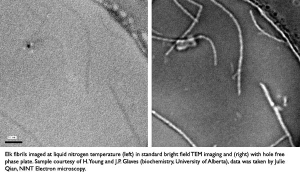
Essentially the simplified phase plate hardware can easily be inserted to optimize the electron optics of the TEM, says Dr. Kawasaki. “It’s very easy to operate compared to other designs where you have to put the beam onto a center of a 1 micron hole. This phase plate has no hole so the beam can be anywhere around the center of a phase plate. There are two places in the column where phase plates can be positioned. One is the objective lens area with the specimen just above. The other area is the selected area aperture (SAA) position which allows more freedom. At NINT, the hole-free phase plates are installed at this position which is much easier to implement and may avoid a contamination issue that may occur under the environment near the specimen.”
Malac added, “You just put the phase plate in the back focal plane and wait for charge to settle down, then take the data.”
Next challenge for the team: investigating now how to control the amount of charge and resulting phase shift.
The two men met in 1999 at Brookhaven National Laboratory, and have worked closely together on developing applications with the TEM at University of Alberta/NINT. Kawasaki consults with advanced researchers using the TEM at labs around the world. As an expert in imaging and analysis, he developed the first S/TEM application for the JEOL TEM in the early 1990s. The function has been essential in nano materials characterization. Malac is a graduate of Charles University in Prague and received his Ph.D. at the University of Alberta.
Phase plates are ideally used to image specimens sensitive to electron beam irradiation: biological materials, protein membranes, and virus particles. Researcher Dr. Holland Chang at University of California (Davis) has used this hole-free phase plate in his recent work.