Inspiring without Overwhelming: Microscopy for K-12 and Some Serious Science
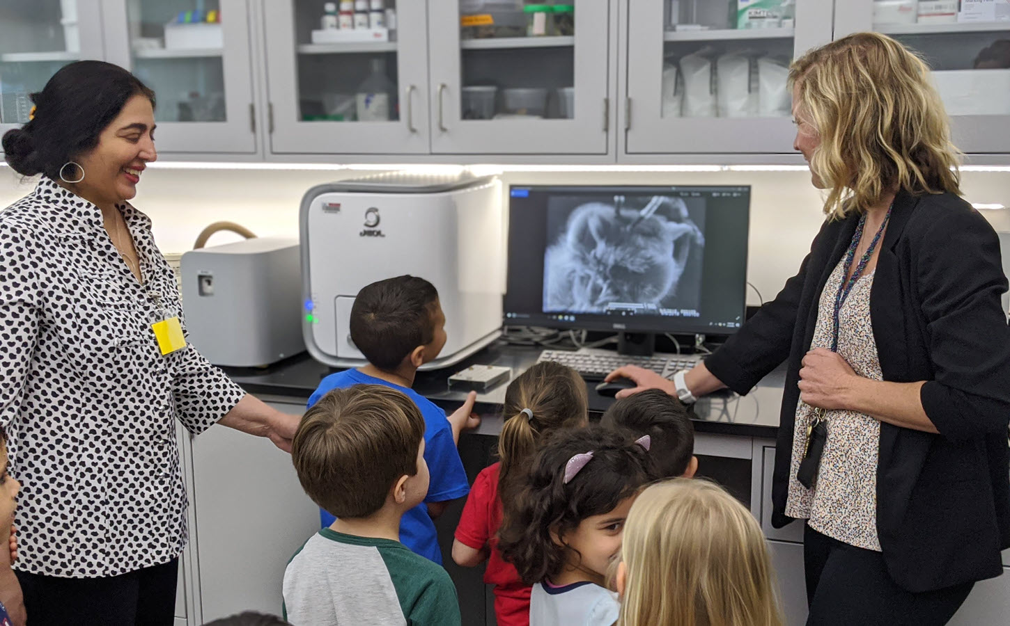
Can you remember the awe of seeing something magnified in a microscope for the first time? That childhood wonder is not lost on the staff at Florida Atlantic University K-12 schools, where they introduce children as young as kindergartners to their NeoScope tabletop Scanning Electron Microscope on their Boca Raton, Florida campus.
“My science heart was just bursting the first time seeing a kindergartner using an electron microscope,” says Dr. Tricia Meredith, Director of Research for Florida Atlantic University’s on-site lab school, A.D. Henderson University School and FAU High School. She is also an Assistant Research Professor in the College of Education at FAU.
Students using the research tools in the FAU High School Owls Imaging Lab began submitting their SEM images to the
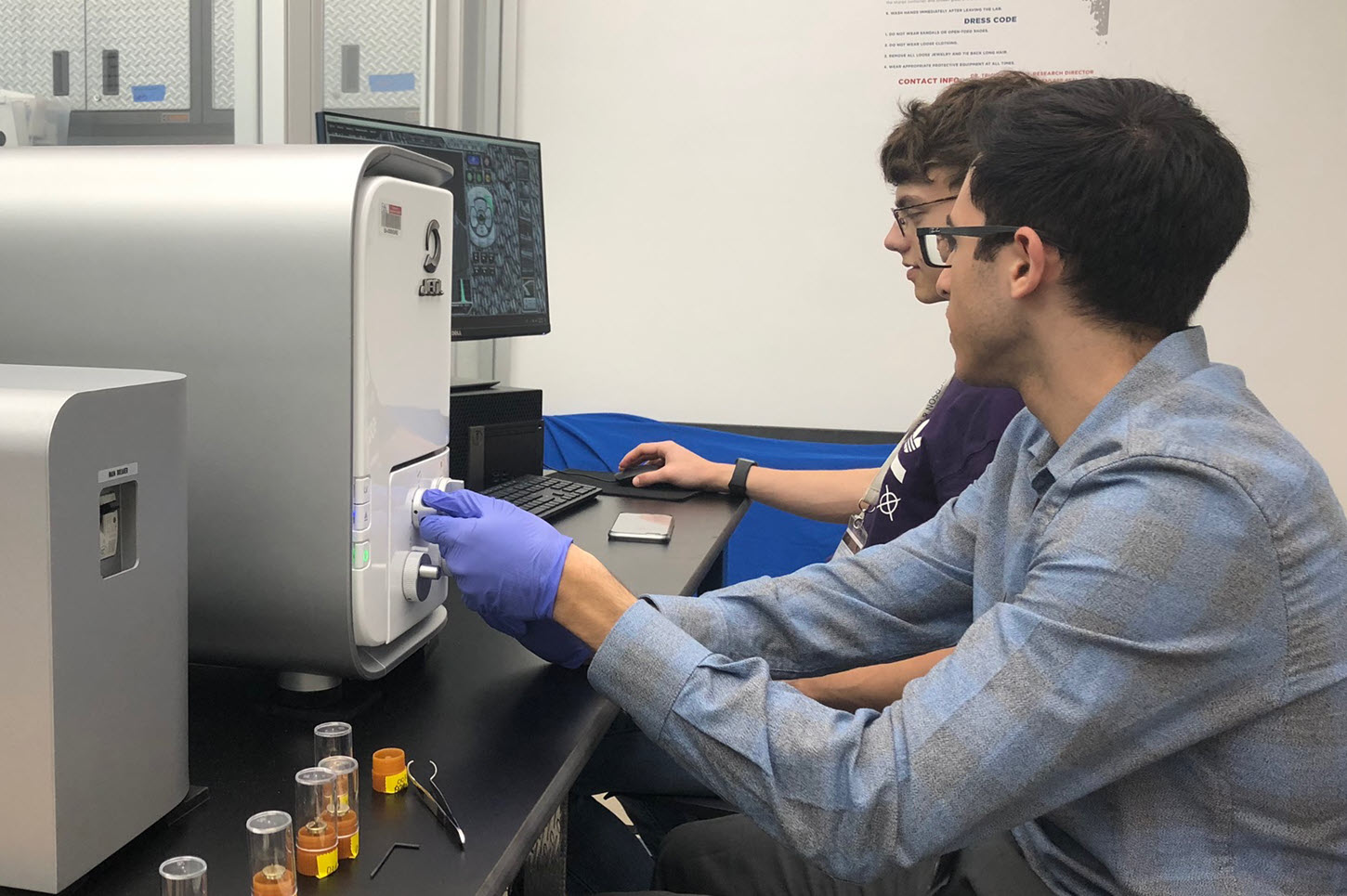
2021 JEOL Image Contest, and they caught our attention with their outstanding results before we realized that they were just kids! (See gallery of images at the top of the page.) With the instruction and guidance of Dr. Meredith, they take images of numerous specimens integral to their research topics in a unique fast-track high school/college program where they complete three years of college requirements after their 9th grade year, which simultaneously satisfy their high school course requirements.
Dr. Meredith manages the Owls Imaging Lab at the school where the goal is two-fold: supporting students in grades k-9 in their early science learning and supporting research and collaborations between students in grades 10-12 and faculty researchers at the university. The SEM is integrated into science classes early on. And the kids learn to do their own work and use the SEM themselves.
“It’s similar to teaching someone how to use a camera,” Dr. Meredith says. “There’s a part that’s training on best practices, then letting them learn by trial and error, then coming in to help them troubleshoot. For example, if they’re getting charging at high mag imaging I’ll say, ‘maybe we need to bring this back to the sputter coater and give it another dusting of gold so we don’t see that’. We do some handholding and a lot of experimentation and trial and error learning. They need the independence to make mistakes.
“The thing I love about the Neoscope is how user-friendly it is. There’s a drawer, three buttons, two knobs, and you click around on the computer monitor until you get the best image. I’m not too afraid they’re going to damage it while I let them play. They can click around and try different specimens and try different settings.”
The SEM is totally accessible to all users, she says. It’s not only a great entry point for even a kindergartner – they get to move the stage around to look at the scales on a butterfly wing – but it also captures the quality of images needed for specific studies that researchers in the university are conducting.
“Our school set up a lab that would be similar to how universities use core facilities. I serve as the PI, but it’s a collaborative space and we give university researchers free access. In exchange they can mentor our undergraduate researchers or do outreach, as is often included in NSF grants.” The students are empowered to act as researchers, collaborating with other, more experienced researchers, and sometimes their early work with the NeoScope prepares them for work with more advanced instruments, like those at FAU’s partner institute, Max Planck Florida Institute for Neuroscience.
Biological samples are the favorite subject in the Owls Imaging Lab, and who better than Dr. Meredith, a biologist herself, to guide students in looking at shark skin, a bee’s hairy eyeball, and gecko toes. Dr. Meredith has conducted research on sharks and their sense of smell, and on the sense of taste using a mouse model. Currently, a university collaborator, Dr. Marianne Porter, is investigating the biomechanics of shark skin and how it can reinforces the body structure during fast swimming. The magnification is suitable for many applications.
Now in her lab she is looking at a variety of wet and conductive specimens requiring sample preparation using a critical point dryer and a sputter coater. Dr. Meredith has learned of a “Nanosuit” product she wants to try for the “squishy, wet specimens.” She has discovered an “untapped love for insects” for the surprise details that students can appreciate. “Plants are really fun, too. The patterns that you find are just so beautiful sometimes.” An image of a rose petal revealing the geometric papules was particularly beautiful.
Student work has also been environmentally conscientious, helping with sea turtle conservation conducted by Loggerhead Marine Life Center. They collected ocean water samples, filtered them down and looked at them in the NeoScope after coating, allowing them to examine and report on marine plankton, bacterium, and microplastics that are in the water.
“I’ve never performed electron microscopy before having the NeoScope,” Dr. Meredith explains. “In a way I wanted something that would be quick and easy enough to learn that it would be accessible for me as the person leading this lab, and for students. I feel I can really connect with the student perspective and their curiosity when you try to inspire without overwhelming them.”
In the weeks that followed our conversation, the second grade students were scheduled to examine their rocks and soil samples in the Owls Imaging Lab, using the stereoscope, electron microscope, and Micro CT scanner to aid them in describing the physical properties. The lab serves as an inspiring resource for students of all ages at FAU.