University of Puerto Rico, Mayagüez Campus Microscopy Center
José R. Almodóvar, Scientific Instrumentation Specialist
When microscopy and photography skills merge, it's possible to achieve art as well as science in a single image. The trained scientist extracts highly detailed information from a sample. But the practiced eye of a professional photographer sees the possibilities of light and shadows, angles, and shapes.
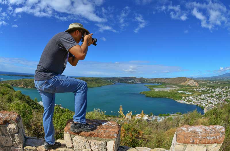
José R. Almodóvar, who is both a biologist and botanist at the University of Puerto Rico, is also an avid nature photographer whose micrographs combine these talents.
 University of Puerto Rico, Mayagüez Campus Microscopy CenterCategory
José R Almodóvar using the SEM: What I saw (and still see) in structural botany – the sub-discipline within botany where my interests lay – was the perfect marriage between the inquisitiveness and exactness that science requires, and the beauty and artistic quality that photography can afford.
 University of Puerto Rico, Mayagüez Campus Microscopy CenterCategory
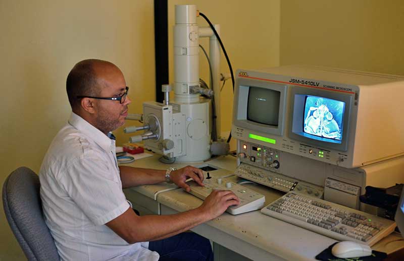
As Scientific Instrumentation Specialist in charge of the operation and maintenance of all the instruments of the Biology Department’s Microscopy Center, at the University of Puerto Rico, Mayagüez Campus, Jose teaches students from various departments. He also images samples for clients from other Universities, agencies and industries who come to the center for structural work. The projects have been tremendously varied, he says, including insect taxonomy, animal reproduction, symbiosis, microbiota, plant taxonomy, morphogenesis, plant development, microbiology, nanotechnology, material science, and agriculture. At present he manages and operates a JEOL JSM-5410LV SEM, JSM-6390 SEM, and a JEM-2100F field emission TEM.
 University of Puerto Rico, Mayagüez Campus Microscopy CenterCategory
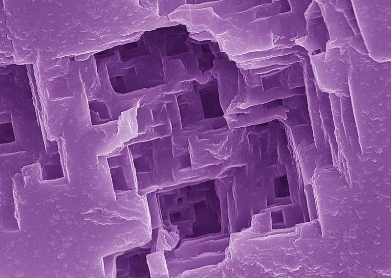
Details in a grain of salt
 University of Puerto Rico, Mayagüez Campus Microscopy CenterCategory
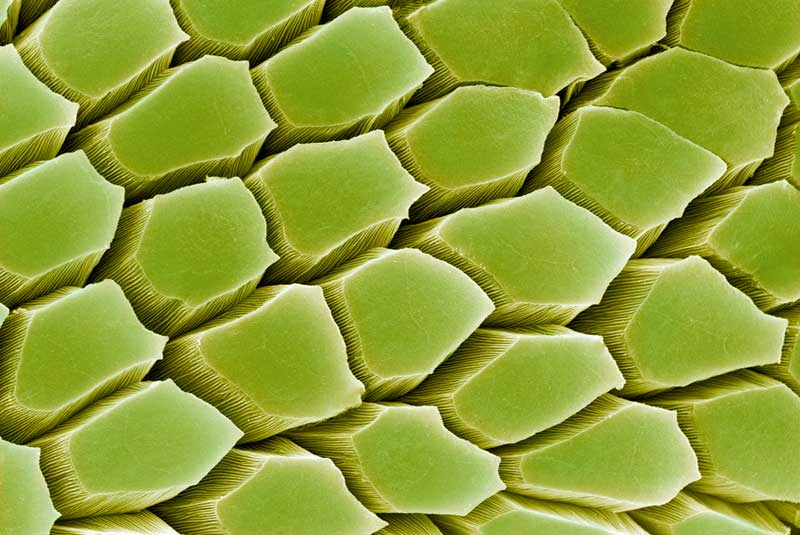 University of Puerto Rico, Mayagüez Campus Microscopy CenterCategory
Tip of the fingers of a coqui (Eleuterodactylus)
 University of Puerto Rico, Mayagüez Campus Microscopy CenterCategory
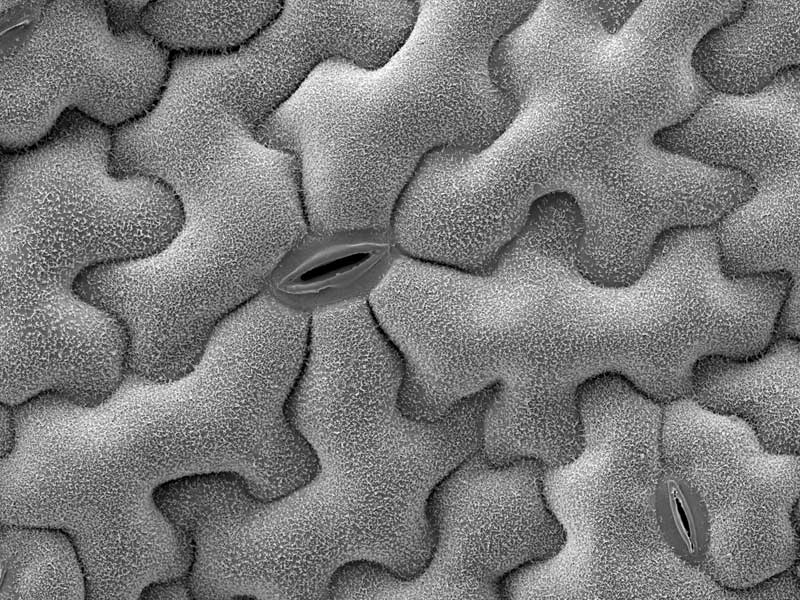
Epidermis and stimata of a leaf from a wild plant
We learned more about Jose's talent for microscopy when he submitted several stunningly beautiful images for the
2014 JEOL Imaging Contest (his image is the April winner).
Which came first for you, photography or microscopy?
I think my passion for microscopy was born when I was an undergraduate student at the University of Puerto Rico, Mayagüez Campus. I remember when I took my first botany course, how I was mesmerized by the unexpected beauty I often found when looking at stained sections of plant tissues. The shapes, patterns, textures and colors where so very reminiscent of art, even though I was undoubtedly studying science.
After completing the General Botany course, I decided to enroll in an undergraduate research course called Special Problems in Botany, under the mentorship of the Biology Department’s structural botanist, Dr. Carlos Muñoz; and I feel in many ways that this experience defined what would become my academic and professional life. The project my mentor assigned me was an anatomical study of Anthurium crenatum (a tropical plant endemic to the area), and I simply fell in love with microscopy and photography. In fact, soon after completing the Anthurium project, I decided that I would pursue a Master’s Degree in Biology to learn more about microscopy and photography.
My Master’s project entailed the anatomical characterization several species of carnivorous plants of Puerto Rico of the genus Utricularia. This project that not only involved a lot of microscopy and microphotography, but it also attracted a lot of attention both, from the academic and general communities. I was even interviewed by a local newspaper that featured an article about my work, which included some of my earliest photographs.
I soon realized that my capacity to collect reliable scientific information depended on my ability to capture images of good quality. And so, as a graduate student at UPRM, I took a Scientific Photography course, which taught me the technical aspects of photography (which still was on acetate film back then). As I gained confidence and experience, I started showing my pictures to more and more people; and it was not uncommon that people would compliment my work and insist that I set up an exhibition, or even compete in photography competitions.
Finally in 2006 I submitted several of my images to Nikon Small World, an international scientific photography competition, and to my surprise and great excitement, one of mine won an award. In retrospect, this was the beginning of my career as a “serious” photographer. I have continued participating in this and other photography competitions, and I feel enormously proud to have received awards almost every single year since.
Do you think being a photographer also helps you create your beautiful SEM images? Could other SEM users benefit from appreciation for photography?
As a photographer, one strives for perfection. As a photographer one learns to see things that most people would overlook. You notice details, you notice distortions, you notice errors. And undoubtedly this obsession to capture the perfect image has helped me to become a better structural biologist as well, since the goal of science is always to get the most accurate representation of reality – without errors, without distortions, with all its details.
What are some of your favorite subjects for both the SLR camera and electron microscope?
My favorite subjects to photograph with the SLR camera are microscopic and small macroscopic organisms, mainly plants, animals, and fungi. The scanning electron microscope is somewhat more restrictive than the camera when choosing suitable subjects to be photographed in that, even at the lowest magnifications, the images are greatly enlarged. Still, some plant parts and very small animals, particularly insects, make great subjects for SEM, and the images obtained can be just as splendid as those captured with a DSLR.
What are your most exciting images so far?
Over the years I have taken so many thousands of pictures that choosing a favorite seems an impossible task (starting with the challenge of remembering them all!). But I can say that many photographs have excited me for many different reasons: some for the fortuitous circumstances that led to capturing them; others for the technical difficulties involved, and the extensive planning and ingenuity required overcome such difficulties; some for their scientific or artistic value; many for the way they have revealed Nature’s magnificent perfection.
One of the most interesting projects ever to come to our Center focused on nanoparticles that would vibrate when exposed to a magnetic field, and which had the potential to be used for the cure of cancer. Another memorable (and extensive) project (it has gone on for over a decade) deals with the interaction between human cells and novel materials for prosthetics.
Do you spend much of your free time in photography, and do you travel much for these photographs?
In all honestly, I am almost addicted to photography… I don’t think it would be too much of an exaggeration to say that, at present, I am spending all of my free time in photography.
I have some done some traveling to capture images that I will always treasure. So far I have gone to Dominican Republic, Mexico, Costa Rica, and several states in the continental United States. I’m hoping to visit many other places!
But the truth is that I do not feel I need to go very far to find incredible photographable subjects. Puerto Rico is a small island with an incredibly rich natural diversity; and this makes me feel incredibly fortunate to live here, as Nature is my favorite subject. Moreover, I have learned that even a potential award-winning photograph might be very, very close, indeed, in the most unexpected place – even in the tiny air bubbles that got accidentally trapped under the cover-slip of the defective microscope slide that I was about to discard…
Also See
University of Puerto Rico Microscopy Center: