 Tubulin Structure
Downing Lab, Lawrence Berkeley National Laboratory
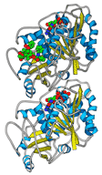
 Dr. Kenneth Downing’s lab at the U.S. Department of Energy Lawrence Berkeley National Laboratory made a major breakthrough for cancer research by solving the atomic level structure of tubulin, the protein responsible for cell division. The team of Eva Nogales, Sharon Wolf, and Kenneth Downing published the paper “Structure of the αβ tubulin dimer by electron crystallography” in Nature in January 1998.
Scientists had been seeking to understand tubulin since the 1950s. Better knowledge of how tubulin polymerizes into filaments that form microtubules and enable a cell to undergo mitosis would open the door to developing new anti-cancer drugs. Already, the natural substance known as Taxol had shown potential in treating a number of cancers by preventing a cell from dividing. It was vital to understand the tubulin structure and how it interacts with Taxol in order to create a more effective drug that targeted only cancer cells.
Downing’s lab was the first to create a 3-dimensional atomic model, and it was achieved through electron crystallography and cryo-EM. The resulting 3D model was a computerized reconstruction of 93 electron diffraction patterns and 159 images culled from more than 4,000 recorded over the previous six years. They were able to work with crystals only one molecule in thickness by obtaining diffraction patterns with an electron beam. In 1998, they achieved 3.7A resolution. Then in 2001, they refined the model to 3.5A resolution and published “Refined structure of alpha-beta tubulin at 3.5A resolution.” Instrumentation for this work includes the JEOL JEM-4000 and the JEM-3100F Transmission Electron Microscopes.